Chest radiographs are frequently performed and a fantastic tool for making diagnoses of acute and chronic conditions as well as acting as a tool for follow-up. If further air accumulates then the air will accumulate lateral and even inferior to the lung.

How To Interpret The Chest X Ray Using A Simple Structured Approach Radiology Student Medical Education Nurse
Example radiograph with left upper zone lesion So you might introduce the above radiograph as follows.

. The x-ray was discovered by German scientist Wilhelm Conrad Röntgen on the 8th of November 1895. Here we can see the pneumothorax within the right apex. Infiltrate on a chest X-ray report is a common finding that radiologists use to describe a white abnormal area of unclear cause.
The interpretation of a chest film requires the understanding of basic principles. In order to identify pneumothorax we need to identify the black air within the pleural space and to differentiate that from the air within the lungs. Chest X-rays is a painless non-invasive test and is the most commonly preferred diagnostic examination to produce images of heart lungs airways blood vessels and the bones of the spine and chest.
The patient will place their chest against a plate which digitally records the image. And interstitial lung pattern appears with diffuse fibrosis. The normal lateral chest x-ray view is obtained with the left chest against the cassette.
The chest radiograph also known as the chest x-ray or CXR is anecdotally thought to be the most frequently-performed radiological investigation globally although no published data is known to corroborate thisUK government statistical data from the NHS in England and Wales shows that the chest radiograph remains consistently the most frequently. AP views of chest X-rays. Frontal chest radiograph of an adult female patient.
Normal anatomy and variants. A chest x-ray is a radiology test that involves exposing the chest briefly to radiation to produce an image of the chest and the internal organs of the chest. Below is a list of terms that radiologists use to describe chest X-rays.
Then find the airway on the x-ray and check to. Systematic approach to the chest film using an inside-out approach. The e-learning resource takes approximately 20 minutes to complete.
If you have symptoms that could be caused. If the x-ray is a true lateral the right ribs are larger due to magnification and usually projected posteriorly to the left ribs Figure-3. In fact every radiologst should be an expert in chest film reading.
The anterior aspect of the 6 th rib the sloping ones should meet the diaphragm near the mid-clavicular line. An abnormal area of infiltrate on a chest X-ray can represent many abnormalities such as infection water or edema tumor abnormal inflammation not related to infection scarring collapsed lung tissue and other things. Note the larger appearing heart on the AP view.
Check adequacy of inspiration. With some of these terms the description can be used to represent several different underlying disease processes while with others the description and disease process are one in the same. Attention should be given to factors such as location size shape and density of an abnormality.
It is for continuous use but can be stopped and restarted at any point. X-rays use high energy rays to take pictures of the inside of your body. It can also check whether a lung cancer has spread.
In the PA view the patient stands with the shoulders rotated anteriorly and depressed hands on the hips. They can show up changes in the lungs. The standard chest X-Rays consists of a PA and lateral chest X-Ray.
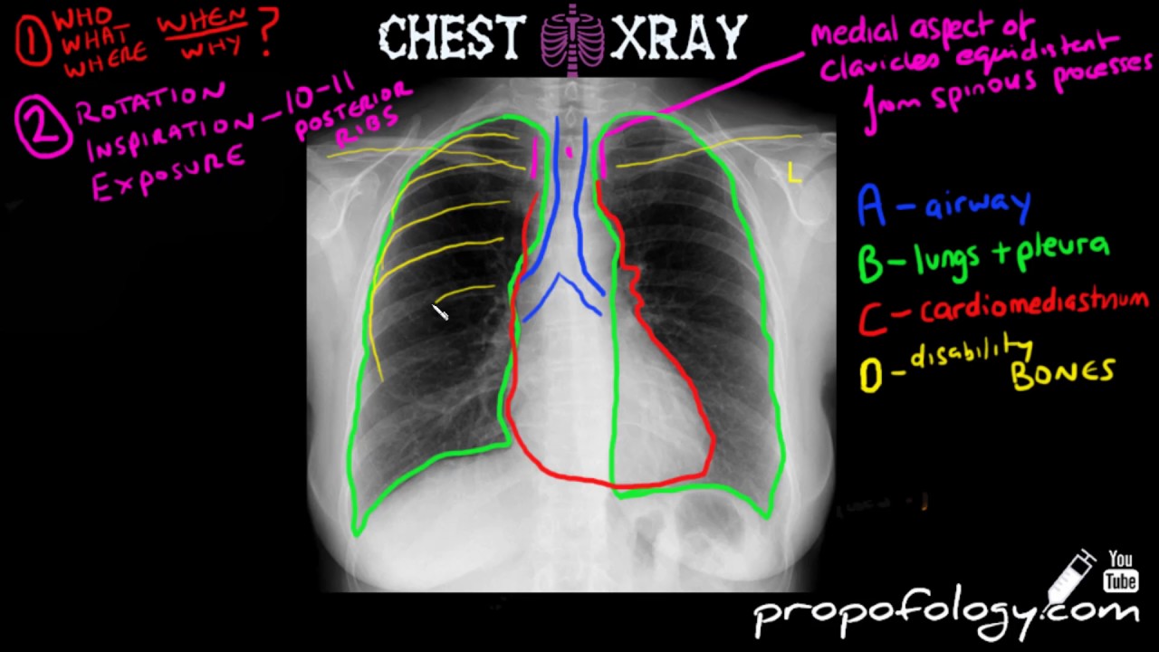
I definitely needed this and I still refer to this method whenever I look at chest x-rays. Technique edit edit source An X-ray uses electromagnetic waves and ionizing radiation to create pictures of the inside of your body. Measure the distance from the medial end of each clavicle to the spinous process of the vertebra at the same level which should be equal.
These terms are often found in a radiology report and are used to describe a process in the lungs. An x-ray exam helps doctors diagnose and treat medical conditions. A chest x-ray produces images of the heart lungs airways blood vessels and the bones of the spine and chest.
X-ray of the chest also known as a chest radiograph is a commonly used imaging study and is the most frequently performed imaging study in the United StatesIt is almost always the first imaging study ordered to evaluate for pathologies of the thorax although further diagnostic imaging laboratory tests and additional physical examinations may be necessary. The chest x-ray is the most frequently requested radiologic examination. Chest X-Ray Views PA and Lateral There are two main views that are used when a chest radiograph is completed.
Comparison of PA vs. X-rays are the oldest and most often used form of medical imaging. The process of description often helps with diagnosis - see the list of lesion descriptors.
Chest x-ray review is a key competency for medical students junior doctors and other allied health professionals. You should be able to see the apices and costophrenic angles. The right ribs red arrows and left.
To read a chest x-ray start by looking for markers on it like L for left R for right PA for posteroanterior and AP for anteroposterior to identify the positioning of the x-ray. It exposes you to a small dose of ionizing radiation to produce pictures of the inside of the body. A chest x-ray is a test that can help to diagnose lung cancer.
These are PA and lateral. Nine pairs of ribs should be seen posteriorly in order to consider a chest x-ray adequate in terms of inspiration. A normal chest x-ray can be used to define and interpret abnormalities of the lungs such as excessive fluid pneumonia bronchitis asthma cysts and cancer.
Changes can be due to cancer but can also be caused by other lung conditions. Article Summary X. Small focal scars can appear as linear densities on a chest x-ray.
This is because the distance is increased between the film and the heartallowing for the X-rays to spread for a greater distance before developing the film Lateral views rightleft. Often a lateral view usually accompanies a PAAP chest X-rayThis can be helpful in settings where the single. It has 19 slides included an initial slide on basics of chest x-ray anatomy a systematic approach on how to review a chest x-ray six common cases a summary table and quiz.
Many are specific to chest X-rays but some are used more generally in radiology. This refers to subtle thin lines and small dots interspersed. Describing a chest X-ray abnormality can be likened to describing a skin rash in a dermatology patient or a lump in a surgical patient.
This is a basic article for medical students and other non-radiologists. This method helps inexperienced students as it reduces the odds to miss anything on a chest x-ray.

Chest X Ray Fundamentals Radiology Medical Coding Radiology Student
How To Read A Chest Xray In 5 Minutes Clinical Youtube X Ray Respiratory Therapy Critical Care
Normal Chest X Ray Medical School Essentials Medical Radiography Medical Student Study

Radiology Chest Xray Normal Radiology Radiology Student Radiology Imaging


0 Comments